

Growth Scans
From 24+ weeks

Growth Scan
An ultrasound in the third trimester may be performed at any stage after 23 weeks, typically around 28-36 weeks gestation. The examination is also know as a growth scan or fetal well-being scan. Scanning at this gestation there is increasing evidence that this allows better detection of growth problems, therefore better outcomes for mum and fetus.
There could be many reasons to have a 3rd trimester ultrasound, this may be due to:
A fetal abnormality was diagnosed on an earlier ultrasound.
That clinically the fetus feels smaller or larger that expected for the gestation.
Small baby in previous pregnancy
History of large for dates or growth restricted in previous pregnancy
Uncertain about the position of the fetal head
Placenta was low-lying on mid trimester scan.
Maternal history; gestational diabetes and high blood pressure
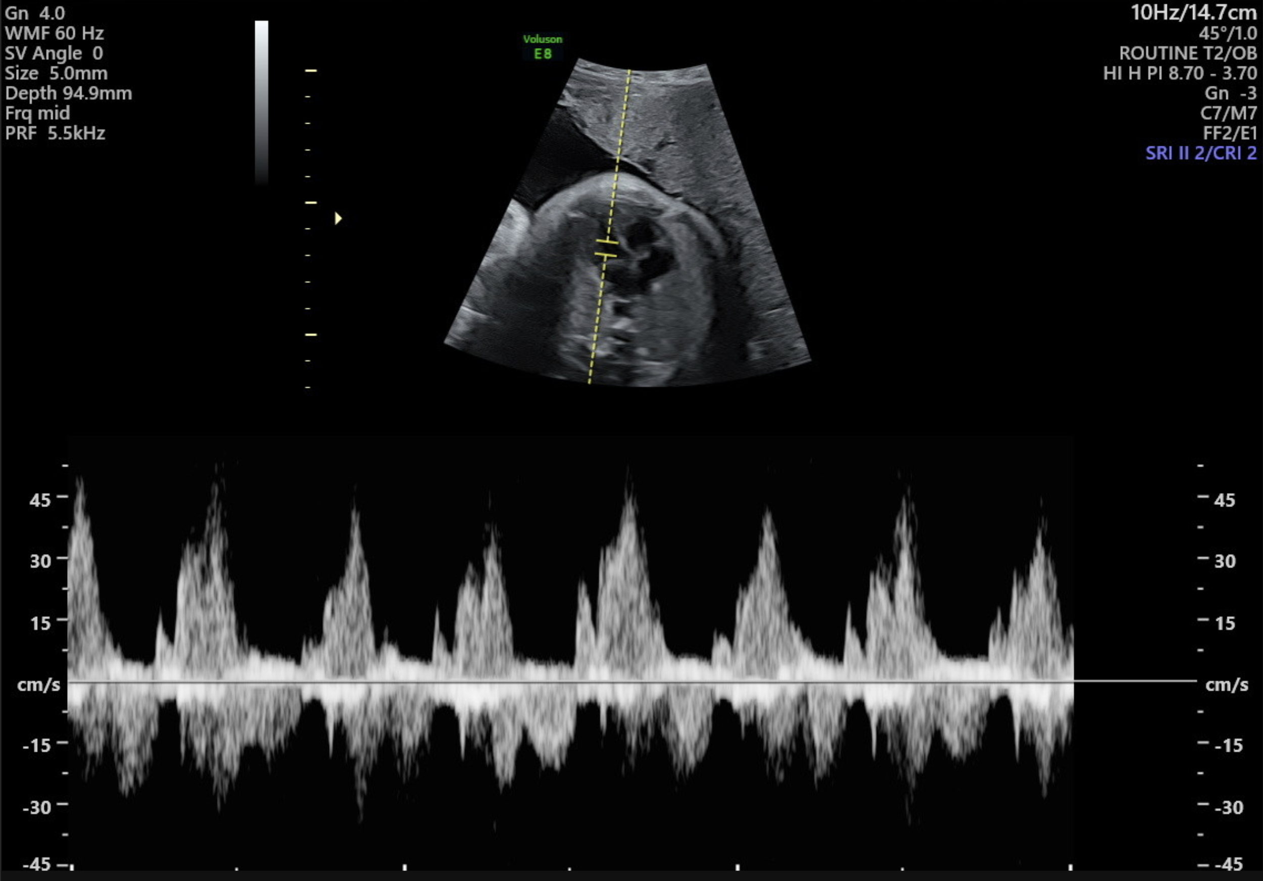
On a 3rd trimester ultrasound the following is examined:
The fetal size and estimated weight. This is calculated from measuring the fetal head, abdomen, femur and humerus. There can be plus or minus 400g on the weight estimate.
The assessment of fetal size/ weight is done by measuring fetal head (Biparital diameter BPD and head circumference HC), abdominal circumference (AC), femur length (FL) and humeral length (HL). The growth of a fetus can only be determined from two separate ultrasounds at least 10 days apart.
Fetal position: Determining whether the fetus is head down ( in the cephalic presentation) or in another position is crucial for planning the delivery.
Fetal anatomy: Although late in the pregnancy the views of certain parts of the fetus maybe difficult. The fetal head (limited assessment), heart, diaphragm, renal pelvis, kidneys, stomach, bladder and cord insertion.
Check for abnormalities. While major structural abnormalities are typically diagnosed in earlier scans, this scan might pick up on any late-developing issues or conditions.
Blood flow in the fetal brain (middle cerebral artery), umbilical cord (umbilical artery) and/or ductus venous when indicated.
Cervical length
Placental position and function. It is essential for the placenta to be in the right location for a safe delivery. The placenta plays a vital role in providing nutrients and oxygen to the fetus. By assessing the blood flow in the umbilical cord and around the placenta, the scan can help to determine whether the fetus is receiving adequate supply of oxygen and nutrients.
Amniotic fluid. The scan can assess the mount of amniotic fluid surrounding the fetus. Adequate amniotic fluid is crucial for protecting the fetus, aids lung development and enabling the fetus to move around. Too much or too little fluid can be a sign of potential problems.
The information from this scan allows the doctor to provide the best possible care for mother and fetus in the later stages of pregnancy and to plan for a safe delivery.
-
To acquire the best diagnostic images possible, the examination is routinely performed Transabdominally, followed by a Transvaginal ultrasound (always with your written consent first).
-
In most cases the examination will be performed transabdominally, but there are some situations where a Transvaginal ultrasound maybe necessary. This improves the assessment of the cervix, placenta and obtain better details of the fetus. In turn this can improve accuracy if the diagnosis.
Transabdominal and transvaginal ultrasound examinations are safe at all stages of pregnancy.
-
The ultrasound procedure typically lasts around 30 minutes. However, the duration may vary depending on the specific reason for the examination and the complexity of the individual case.
-
If possible we recommend wearing comfortable, loose-fitting clothing that provides easy access to the area being imaged. Two-piece clothing, with separate upper and lower garments, is preferable.
Additionally, please ensure that you empty your bladder 1 hour before the procedure and then drink 2 glasses (600ml) of water, holding it without emptying your bladder again. Please note that your appointment might be delayed if your bladder is not adequately full.
-
Having some urine in your bladder can be beneficial for outlining the cervix and visualizing the relationship of the placenta with the lower uterus during imaging. It also helps to elevate the uterus from the pelvis into the abdomen, facilitating better visualization of the fetus.
You don't need to have a large amount of urine, and simply drinking a glass of water 30-60 minutes before your scheduled appointment is sufficient.
-
We welcome one adult support person, whether it be your partner or a family member, to accompany you during your Diagnostic scan. However, for specific reasons, we have implemented policies that prohibit children from attending Diagnostic ultrasound appointments. While we understand that this may be disappointing for families, it is essential to maintain a focused environment. Thus allowing the Sonographer to be able to concentrate during the scan whilst taking the necessary images and measurements. Occasionally, our sonographer may need to discuss unfortunate news found during your appointment and so this must be taken into consideration. The presence of children can introduce distractions that may impact the quality of the examination.
-
At Trinity Imaging, we provide our patients with their images to an app on your phone called Tricify. For further details please ask our receptionist at your appointment.


All our scans are performed in our premium-furnished luxury viewing room specially designed for you.
We are all looking forward to getting to know you, your baby and family along your pregnancy journey.
Our Growth Scans take up to 30 minutes and include:
2 x 3D thermal printed hard copy images
All pictures taken on the day straight to your mobile phone